
Dr. Friedlander's
Contributions to Neuroscience





Videos of neurons in culture undergoing programmed cell death. Understanding how neurons die provides a substrate to study the process to develop therapies for neurological diseases featuring neuronal death.
Dr. Friedlander’s scientific work helped open the field of neurotherapeutics by showing that neurodegeneration and acute central nervous system injury are driven, at least in part, by discrete pathogenic pathways that can be mechanistically defined and therapeutically manipulated. Across chronic neurodegenerative disorders such as Huntington’s disease and amyotrophic lateral sclerosis (ALS), and across acute neurologic injury including ischemic stroke, traumatic brain injury, and spinal cord injury, Dr. Friedlander’s research established that these conditions are not simply descriptive clinicopathologic syndromes, but biologically tractable processes in which targeted intervention can slow disease progression, reduce tissue injury, and preserve neurologic function.
In foundational work carried out while Dr. Friedlander was a postdoctoral fellow in Dr. Junying Yuan’s laboratory, they showed that dominant-negative inhibition of ICE/caspase-1 protects neurons from trophic factor withdrawal and reduces ischemic brain injury, helping establish the first functional role of a caspase pathway in neurologic disease (Friedlander et al., J Exp Med, 1997). They then extended this framework to ALS, showing that neuronal expression of a dominant-negative Caspase-1 inhibitor slowed symptomatic progression and delayed mortality in mutant SOD1 mice, the first demonstration that genetic manipulation could slow disease progression in ALS (Friedlander et al., Nature, 1997).
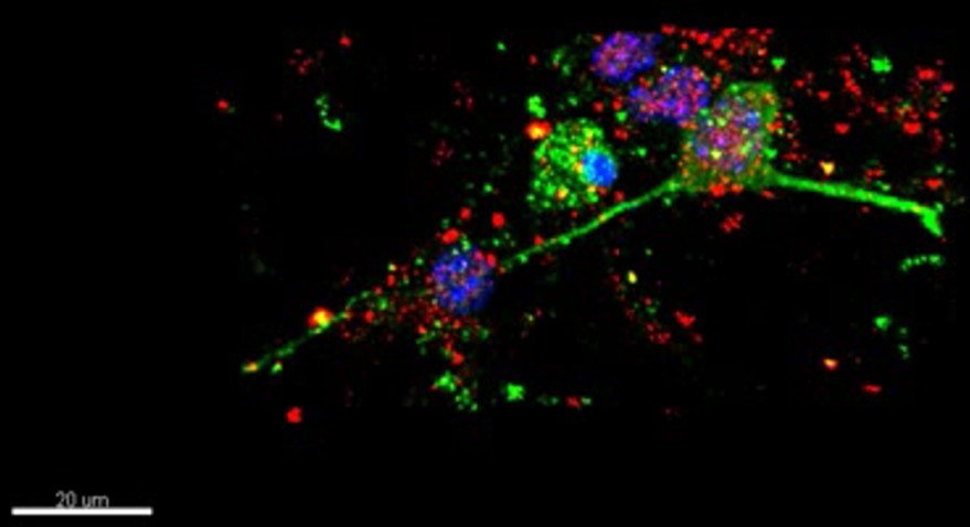
Neurons were subjected to OGF then fixed and stained with Active Caspase-3 and CytC antibodies. DAPI (nucleus).
Dr. Friedlander’s laboratory at the Brigham and Women’s Hospital/Harvard Medical School next demonstrated that the same mechanistic logic could be applied to Huntingto’s disease (HD). His group showed that caspase-1 is activated in mouse and human HD brain and that dominant-negative caspase-1 as well as intracerebroventricular delivery of caspase inhibitors delayed symptom onset and extended survival in the R6/2 model—the first demonstration that disease progression and mortality could be slowed in HD by genetic and pharmacologic manipulation (Ona et al., Nature, 1999). This therapeutic principle was then reinforced by showing that minocycline delayed progression and mortality in HD mice (Chen et al., Nature Medicine, 2000). In ALS, Dr. Friedlander and colleagues extended pathway-based therapy pharmacologically by demonstrating that broad caspase inhibition delayed onset and prolonged survival (Li et al., Science, 2000) and that minocycline delayed onset and extended survival while identifying inhibition of mitochondrial cytochrome c release as a central protective mechanism of action of minocycline (Zhu et al., Nature, 2002). Together, these studies established that disease progression in major neurodegenerative disorders could be altered through rational intervention in defined pathogenic pathways.
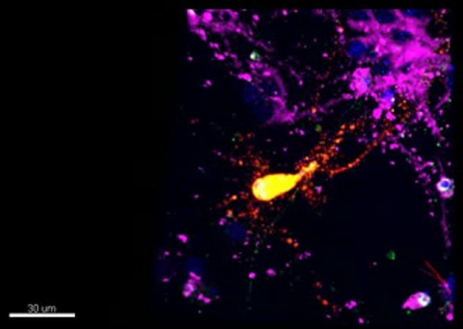
Dead neuron transfected with β-Gal and stained against active Caspase-3 and preloaded with Mitotracker. Nucleus stained with DAPI. Fixed sample.
The same principles were extended to acute neurologic injury. In traumatic brain injury, Dr. Friedlander and colleagues showed that caspase-1 is activated after brain trauma and that genetic and pharmacologic inhibition of the caspase-1 cascade reduces tissue injury and free-radical production (Fink et al., Neuroscience, 1999). They then demonstrated neuronal localization of activated caspase-1 and caspase-3 after traumatic brain injury and showed that minocycline reduces lesion volume, caspase-1 activity, and neurologic dysfunction (Sanchez Mejia et al., Neurosurgery, 2001). In spinal cord injury, Dr. Friedlander’s laboratory demonstrated a functional role for neuronal caspase-1 and caspase-3 in traumatic SCI (Li et al., Neuroscience, 2000) and later showed that minocycline inhibits contusion-triggered mitochondrial cytochrome c release, spares tissue, and improves long-term functional recovery (Teng et al., PNAS, 2004). These studies helped define a unifying principle of acute neuroprotection: that injury-activated cell-death pathways are modifiable therapeutic targets rather than inevitable downstream consequences of damage.
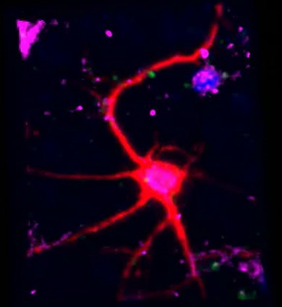
Healthy neuron transfected with β-Gal and stained against active Caspase-3 and preloaded with Mitotracker. Nucleus stained with DAPI. Fixed sample.
In later work, Dr. Friedlander made major contributions to mitochondrial neurobiology in Huntington’s disease and aging-related neurodegeneration. His laboratory demonstrated that mutant huntingtin directly targets the TIM23 mitochondrial protein import machinery and impairs protein import in synaptosomal mitochondria early in Huntington’s disease, identifying mitochondrial import failure as an early pathogenic event rather than a late epiphenomenon (Yano et al., Nature Neuroscience, 2014). Dr. Friedlander’s group then showed that melatonin is synthesized within the neuronal mitochondrial matrix and signals through a mitochondrial MT1 GPCR pathway to block cytochrome c release and caspase activation, revealing an automitocrine intrinsic mitochondrial neuroprotective system (Suofu et al., PNAS, 2017). Building on that discovery, Dr. Friedlander and colleagues demonstrated that melatonin depletion associated with aging and neurodegeneration promotes mitochondrial DNA release and cGAS/STING/IRF3-driven neuroinflammatory signaling, thereby linking declining mitochondrial melatonin to neuronal and synaptic vulnerability in aging and neurodegeneration (Jauhari et al., JCI, 2020).
Together, these discoveries helped establish a modern therapeutic view of neurodegeneration in which caspase activation, mitochondrial dysfunction, and neuroinflammatory signaling are not merely correlates of disease, but actionable drivers of it. That body of work constitutes Dr. Friedlander’s scientific legacy.