
Arteriovenous Malformations (AVMs)
AVMs require careful evaluation to determine whether treatment is appropriate and, if so, which approach is most suitable.
Management may involve surgery, embolization, radiosurgery, or a combination. The goal is to develop a strategy that balances effectiveness with safety, based on the specific characteristics of the lesion and the patient.

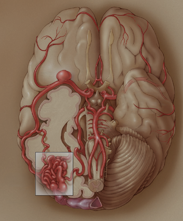
What Is a Brain AVM?
An arteriovenous malformation — or AVM — is an abnormal tangle of blood vessels in the brain that disrupts the way blood normally flows. In a healthy brain, arteries bring oxygen-rich blood to brain tissue, and veins carry it back out. In an AVM, vessels form a chaotic knot that creates a direct, high-pressure shortcut between arteries and veins, bypassing the surrounding brain entirely.
This abnormal routing puts increased stress on the vessel walls over time. If those walls weaken and rupture, it can cause bleeding in the brain — which is why AVMs, even when silent, deserve careful expert evaluation.
AVMs are typically present from birth. They are not caused by anything the patient did, and in most cases they are not inherited.
Surgical images are shown below for educational purposes.
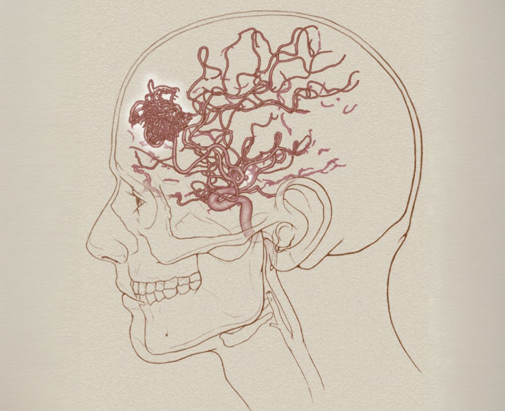
An AVM (shown in red) is a tangle of abnormal vessels that diverts blood away from healthy brain tissue.
Presenting Symptoms
A brain AVM may not cause symptoms for many years and may be discovered incidentally on imaging done for another reason. When symptoms do occur, they most commonly result from either bleeding or seizures.
The symptoms at presentation depend on the location of the AVM and the area of brain involved. When an AVM bleeds, the most common symptoms are sudden headache and neurologic deficits related to the location and size of the hemorrhage. Similarly, the type of seizure often reflects where the AVM is located in the brain.
New symptoms may include:
- A first seizure
- Sudden or severe headache
- Weakness, numbness, or tingling
- Vision changes
- Difficulty speaking or understanding language
Other new neurologic symptoms should be evaluated by a neurologist or neurosurgeon and often require brain imaging.
AVMs can also occur alongside other vascular abnormalities. In some patients, a flow-related aneurysm forms on one of the arteries that feeds the AVM. This finding can significantly influence the treatment plan and should be considered as part of the overall care strategy.
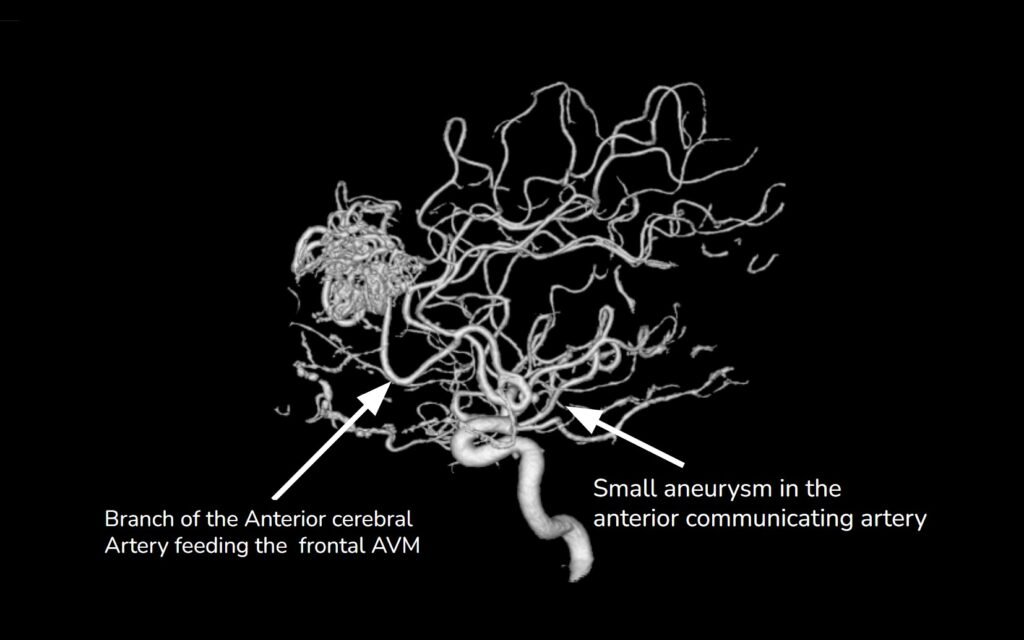
An angiogram showing a branch of the anterior cerebral artery feeding a frontal AVM. A small aneurysm (indicated) is also visible on a nearby vessel — a common finding in some AVM patients that requires treatment.
How an AVM Is Diagnosed & Graded
When an AVM is suspected, Dr. Friedlander uses a combination of imaging studies to build a complete picture of the malformation — its size, location, and the blood vessels that supply and drain it. This information guides every decision about treatment.
CT scan: Often the first study performed, especially when a patient presents with a seizure or sudden symptoms. It can detect bleeding and give an immediate view of the AVM’s location in the brain.
MRI: Provides detailed images of the AVM and the brain tissue around it. MRI is particularly useful for identifying whether the AVM is near critical areas responsible for movement, speech, or vision.
Cerebral angiography: The gold standard for AVM evaluation. By injecting contrast dye into the vessels, angiography maps exactly which arteries are feeding the AVM and how blood drains away from it — information that is essential for surgical planning and cannot be obtained any other way.
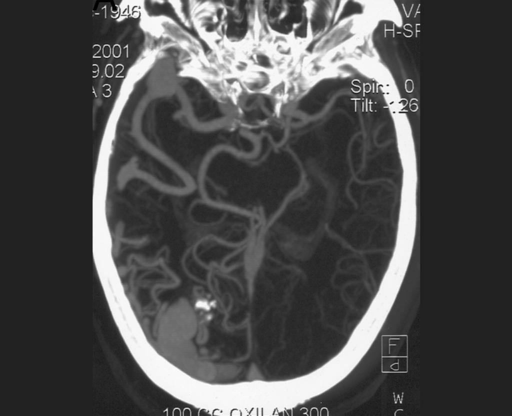
Pictured above is the CT scan of a patient who presented with a seizure and was neurologically intact, with no significant past medical history. Imaging showed no evidence of acute intracranial hemorrhage.
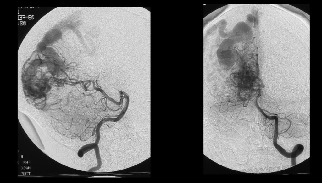
A cerebral angiogram showing the AVM and the network of blood vessels surrounding it. This detailed imaging helps Dr. Friedlander map exactly where the malformation is and how blood is flowing through it.
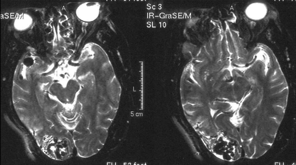
Two MRI sequences of the same patient, viewed side by side. Different imaging settings highlight different features of the AVM and its relationship to surrounding brain tissue, helping the surgical team plan the safest possible approach.
Understanding the Spetzler-Martin Grade
Once imaging is complete, Dr. Friedlander assigns a Spetzler-Martin grade — a standardized score used by neurosurgeons worldwide to assess the complexity of an AVM and the risk of treatment. Three factors determine the grade:
- How large the AVM is
- Whether it sits in or near an “eloquent” region — a brain area responsible for essential functions like movement or speech
- How the AVM drains — whether its veins run toward the brain’s surface or deep into the brain
Grades run from I (lowest complexity) to V (highest). A higher grade signals a more complex lesion.
Patient Case Example
A 51-year-old woman came to Dr. Friedlander after experiencing a seizure. She was neurologically intact — no weakness, no speech difficulty, no deficits of any kind. A CT scan showed no evidence of bleeding. Imaging revealed an occipital AVM fed by a branch of the middle and posterior cerebral arteries, with a feeding artery irregular aneurysm.
Her AVM was classified as Spetzler-Martin Grade III — scored on size, location near functional brain tissue, and a favorable venous drainage pattern. Dr. Friedlander designed a staged, three-part treatment plan: first, surgical clipping of the aneurysm to eliminate its risk of rupture; next, embolization of the main artery feeding the AVM to reduce blood flow into it; and finally, microsurgical resection to remove the malformation completely. Post-operative imaging confirmed the AVM was entirely gone.
View treatment below
Treatment: A Staged, Personalized Approach
Treating a complex AVM often requires more than one procedure. For this patient — and for many others with Grade III or higher lesions — Dr. Friedlander used a carefully sequenced plan designed to balance complete AVM removal with preservation of neurological function.
Step 1 — Clipping the aneurysm Because an aneurysm was present on a vessel feeding the AVM, it was treated first. A small titanium clip was placed at its base to seal it off permanently, reducing its risk of rupture before addressing the AVM.
Step 2 — Embolization Before surgery, blood supply to the AVM was reduced by precisely injecting a material which selectively blocks flow using a procedure called embolization. A thin catheter was guided through the bloodstream to the feeding vessel, where a special material was injected to reduce blood flow into the AVM. Less blood flow during surgery means less risk of bleeding and a more controlled resection.
Step 3 — Microsurgical resection With the blood supply reduced, Dr. Friedlander surgically removed the AVM under a high-powered microscope. Working with careful microsurgical planning and technique through the natural spaces of the brain, he separated the tangled vessels from surrounding healthy tissue and removed the malformation entirely. The goal — and the result — was complete removal.
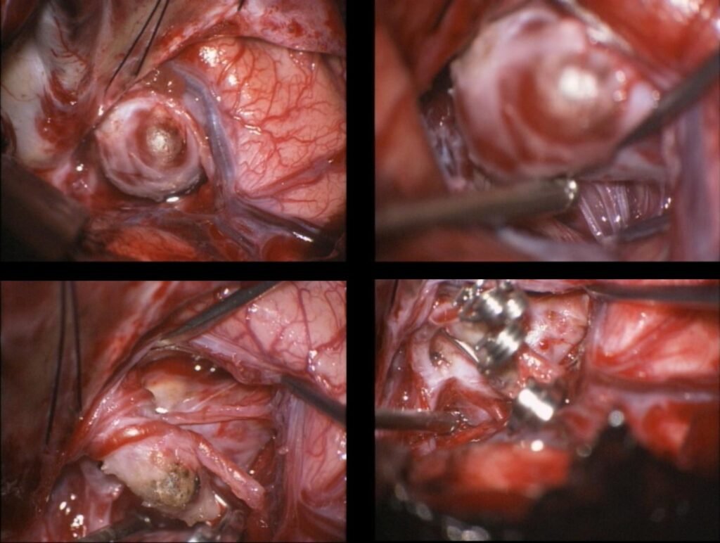
Intraoperative views showing the aneurysm with the titanium clips applied.
After Surgery: Confirming the AVM Is Gone
After AVM resection, complete removal must be confirmed — not assumed. Even a small remnant can continue to carry a risk of bleeding. When surgery is appropriate, the goal is complete AVM removal while minimizing neurological risk.
Following surgery, angiography is performed to verify that the AVM has been completely eliminated. This imaging is the definitive proof of success.
For patients treated for an unruptured AVM, recovery is typically smooth. Most return to their normal activities within weeks, with follow-up imaging scheduled over the coming months.
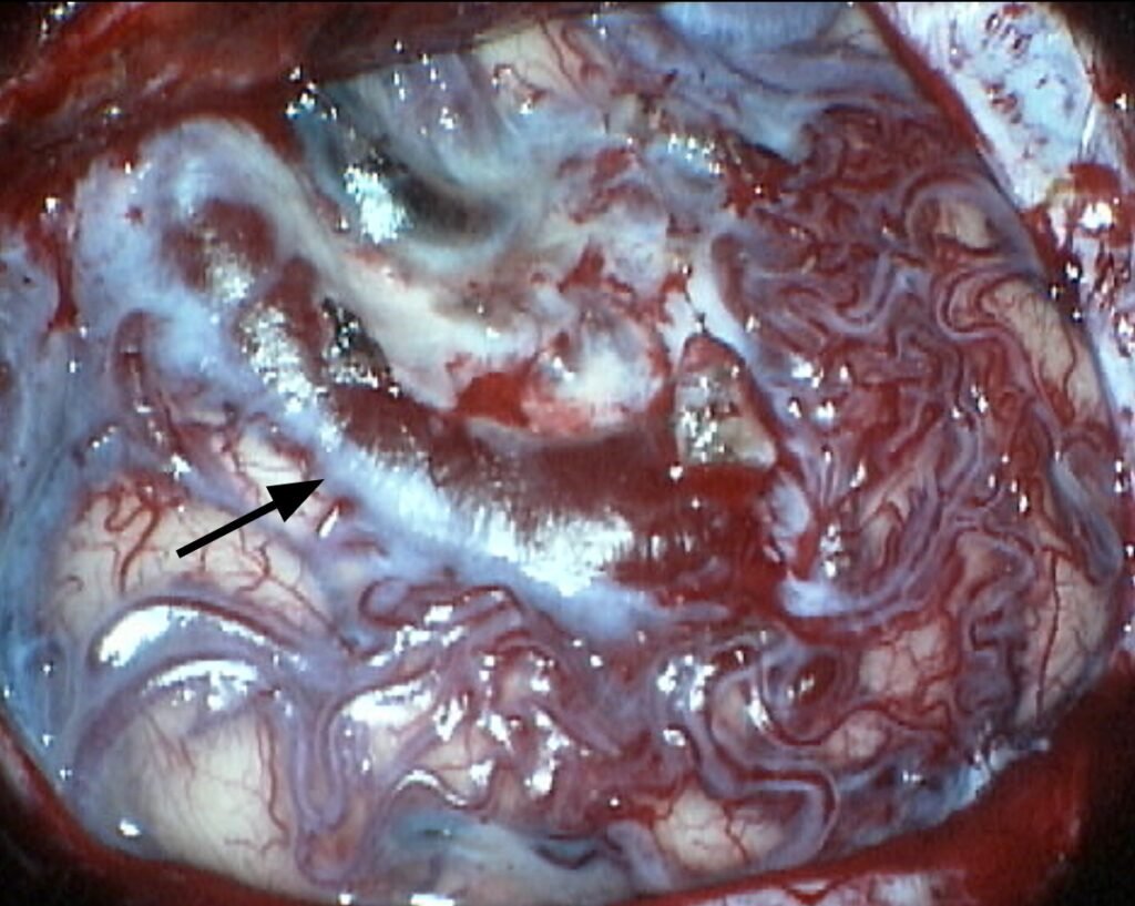
An intraoperative view of the AVM through the surgical microscope. The dense tangle of abnormal vessels is clearly visible. The arrow points to the main draining vein.

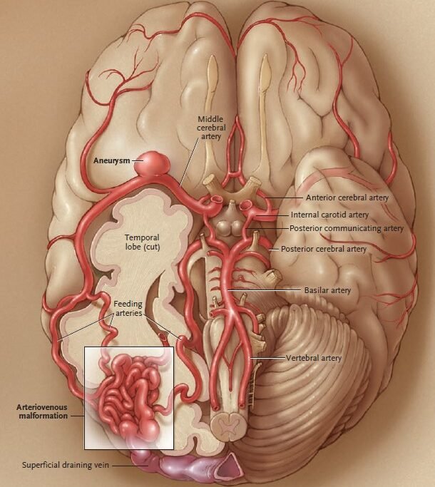
Further Reading: AVMs in the New England Journal of Medicine
For a deeper look at the diagnosis and treatment of brain arteriovenous malformations (AVMs), explore Dr. Friedlander’s peer-reviewed article published in the New England Journal of Medicine.

Request a Consultation with
Dr. Friedlander
dr.rf@iss.org
Phone
845·764·4353
Address