
Brain Tumors
The key decisions in brain tumor care are whether surgery is indicated and what the goals of surgery should be.
These decisions require balancing tumor control with preservation of neurological function. This balance is individualized and depends on tumor type, location, and the patient’s condition.
Dr. Friedlander works closely with each patient to define a clear and appropriate strategy, with an emphasis on experience-based judgment and precision in execution.

Understanding Brain Tumors
Brain tumors vary widely in type, size, and location. About 85% of meningiomas — one of the most common brain tumor types — are benign. Surgery may be recommended when a tumor is causing symptoms such as:
- Neurological deficits or weakness
- Memory or cognitive changes
- Visual loss
- Seizures
- Gait instability or balance problems
- Headaches from pressure on the brain
A thorough evaluation considers your age, the tumor’s size and growth, and your overall health before any decision is made.
Surgical images are shown below for educational purposes.
Types of Tumors Dr. Friedlander Treats
Meningioma
Meningiomas grow from the lining of the brain. They are usually benign and slow-growing, but their location — deep near the brainstem, within the brain’s fluid spaces, or spread across the frontal region — determines the complexity of treatment.
Gliomas
Gliomas, and especially glioblastoma (GBM), are among the most complex and aggressive brain tumors. Their treatment requires experience, careful judgment, and a tailored approach that considers each patient’s tumor location, imaging features, neurologic function, overall health, and goals of care.
Surgery focuses on removing as much tumor as safely possible while protecting surrounding healthy brain, particularly areas responsible for speech, movement, cognition, and other essential neurologic functions. Achieving this balance requires meticulous planning, advanced surgical tools, and the experience to define and pursue the safest surgical goals for each individual patient.
Spinal Cord Tumors
Tumors of the spinal cord can cause pain, weakness, or numbness that travels down the arms or legs. Surgical removal follows the same principles of precision and minimal disruption to healthy tissue.
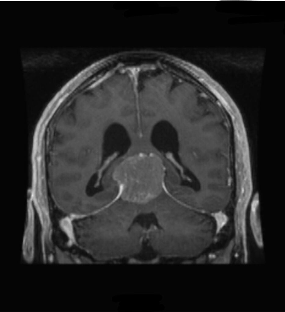
5 cm deep Anterior Tentorial Meningioma.
Surgical Approach
Dr. Friedlander’s principle is simple: to achieve the appropriate extent of tumor removal while preserving neurological function.
Every surgery begins long before the operating room. Using advanced imaging including tractography — a technique that maps the brain’s critical nerve pathways in three dimensions — we plan each procedure to remove the tumor while protecting everything around it.
During surgery, we focus on:
- Protecting veins, arteries, and healthy brain tissue at every step
- Reducing brain swelling before and during the procedure
- Achieving complete removal wherever safely possible
- Monitoring brain function in real time
No two tumors are alike, and no two surgical plans are identical.
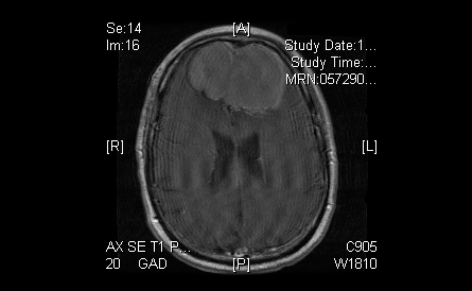
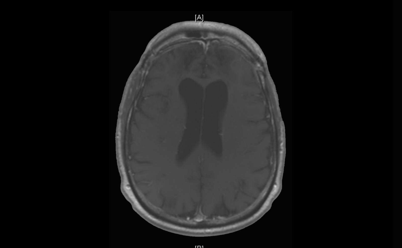

The 8 cm bifrontal meningioma shown here was completely removed using this approach.
Meningioma
Meningiomas are the most common brain tumor Dr. Friedlander treats. They grow from the meninges — the protective lining that surrounds the brain and spinal cord. The vast majority are benign, and many grow slowly over years before causing symptoms.
Because meningiomas can grow in many different locations, the symptoms they cause and the surgical approach required vary greatly. For many meningiomas, the goal of surgery is complete removal when it can be achieved safely. In others, preservation of neurological function may appropriately take priority over complete resection.
Surgery may be recommended when the tumor is causing:
- Neurological deficits such as weakness, numbness, or vision loss
- New and different headaches, particularly morning headaches.
- Cognitive changes or memory difficulties
- Seizures
- Gait instability or balance problems
- Significant brain swelling
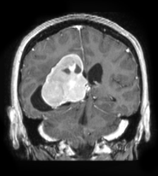
MRI of a 57 yo female with right visual loss.
Meningiomas are classified by where they grow:
Anterior Skull Base / Frontal Tumors
These tumors at the front of the skull base can grow large before causing noticeable symptoms. A bicoronal incision — a carefully placed forehead incision — allows full access to these tumors while leaving a well-hidden scar. Even an 8 cm bifrontal meningioma can be completely removed using this approach.
Tentorial
The tentorium is a fold of tissue deep inside the skull that separates the upper and lower brain. Meningiomas that grow here sit close to the brainstem — the structure that controls breathing, consciousness, and basic body function. Surgical removal requires exceptional precision.
Intraventricular
The ventricles are fluid-filled spaces deep within the brain. Tumors that grow here — sometimes reaching 7 cm or larger — require navigating deep into the brain, minimizing disruption to surrounding tissue. Complete removal is the goal, and outcomes are monitored over time with follow-up imaging.
Clinoidal and Sphenoid Wing
These tumors grow near the base of the skull, close to critical arteries and the optic nerve. Vision loss is a common presenting symptom. Surgical planning must account for the proximity of these vital structures.
Convexity and Superior Sagittal Sinus
Convexity meningiomas grow on the outer surface of the brain and are often more accessible, though size and involvement of major veins — particularly the superior sagittal sinus — can complicate removal. Venous preservation is a priority.
Special Considerations for Older Patients
Meningiomas are more common in older adults, and age alone is never a reason to avoid treatment. Surgical planning for elderly patients takes into account overall health, tumor behavior, and the balance between the risks of surgery and the risks of leaving the tumor untreated.
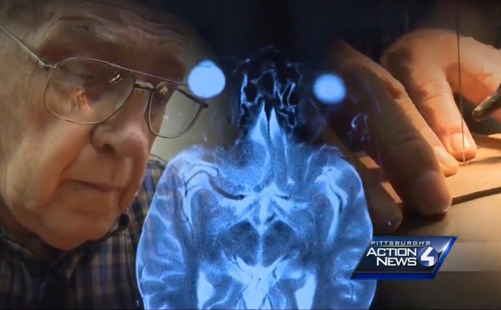
A Patient Story: John Fisher, Woodworker
John Fisher had spent his life creating with his hands — until a benign tumor pressing on his optic nerve threatened his vision and independence.
Without treatment, he risked losing both his sight and cognitive function.
“I hate to say it: I didn’t want to live.”
Dr. Friedlander performed a frontotemporal craniotomy with orbital osteotomy, safely removing the tumor while protecting surrounding brain tissue. John regained his full vision.
“Every time I think about this, I think about how lucky I am.”
Glioblastoma (GBM)
Glioblastoma is the most aggressive primary brain tumor. Unlike meningiomas, GBM infiltrates brain tissue rather than displacing it, which makes complete surgical removal impossible — but maximum safe resection significantly improves outcomes and extends the effectiveness of follow-up treatment.
Surgery for GBM is guided by advanced imaging at every stage:
- Pre-operative tractography maps the brain’s critical white matter pathways — the highways that carry signals for movement, language, and vision— helping plan a surgical approach designed to minimize injury to these pathways.
- Post-operative imaging confirms the extent of resection and guides the next steps in care
After surgery, treatment continues with radiation oncology and neuro-oncology as part of a coordinated multidisciplinary plan.
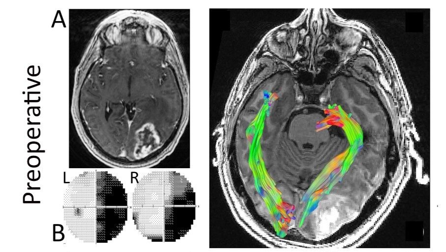
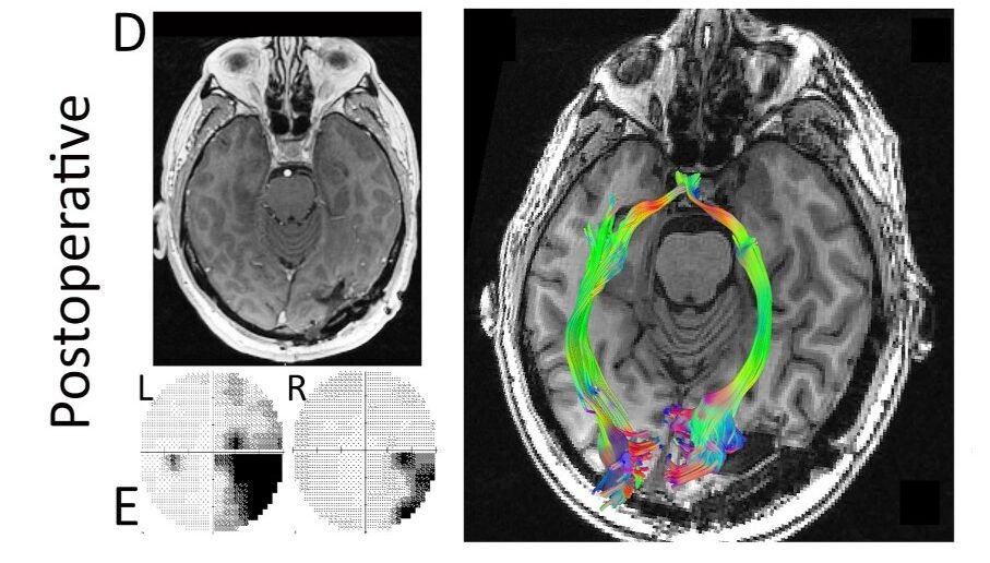
MRI, visual field test, and tractography results of a patient pre-op and post-op.
Spinal Cord Tumors
Tumors of the spinal cord are less common than brain tumors but carry their own set of challenges. They can be located inside the spinal cord itself (intramedullary), within the surrounding membrane (intradural extramedullary), or outside the dura. Each location calls for a different surgical strategy.
Symptoms often include:
- Pain that radiates down the arms or legs
- Progressive weakness or numbness
- Loss of coordination
- In severe cases, bowel or bladder dysfunction
The same principles that guide brain tumor surgery apply here — precise microsurgical technique, careful preservation of surrounding neural tissue, and a goal of complete removal wherever safely achievable. Intraoperative monitoring of spinal cord function is used throughout the procedure to protect neurological function in real time.
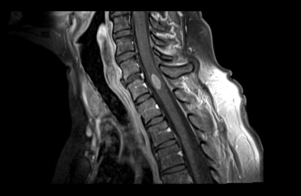
MRI of a spinal cord tumor.
Request a Consultation with
Dr. Friedlander
dr.rf@iss.org
Phone
845·764·4353
Address