
Brain Aneurysm
For patients with an unruptured aneurysm, the central question is whether treatment is necessary.
This requires careful assessment of both patient-specific and aneurysm-specific factors. The goal is to weigh the risks of observation against the risks of intervention in a clear and individualized way.
When treatment is indicated, options are considered thoughtfully, including both microsurgical and endovascular approaches.

What is a Brain Aneurysm?
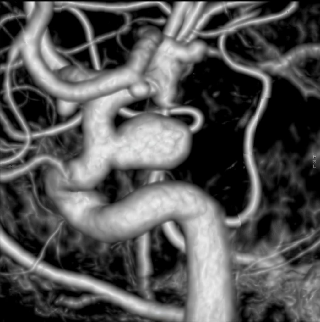
A brain aneurysm is a weak or thin spot on a blood vessel in the brain that balloons outward and fills with blood. Over time, the pressure of blood flowing through can cause this bulge to grow — and in some cases, to rupture. While the word “aneurysm” can feel frightening, many are found incidentally and treated successfully before they ever cause a problem.
Dr. Friedlander is a nationally recognized expert in the surgical treatment of brain aneurysms, with decades of experience helping patients understand their options and select the treatment strategy best suited to the aneurysm and the individual patient.
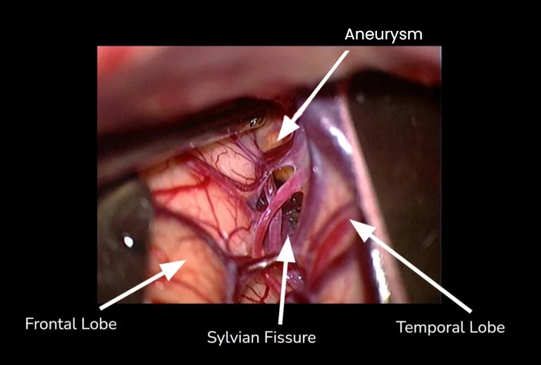
Surgical images are shown below for educational purposes.
Causes, Symptoms & Risk Factors
Most brain aneurysms develop over time without a clear cause or predisposition. However, they are also more common in association with genetic, medical, and lifestyle factors.
You may be at higher risk if you:
- Have one or more first-degree relatives who have had a brain aneurysm
- Have high blood pressure (hypertension)
- Smoke or have smoked
- Have a connective tissue disorder such as Marfan syndrome or Ehlers-Danlos syndrome
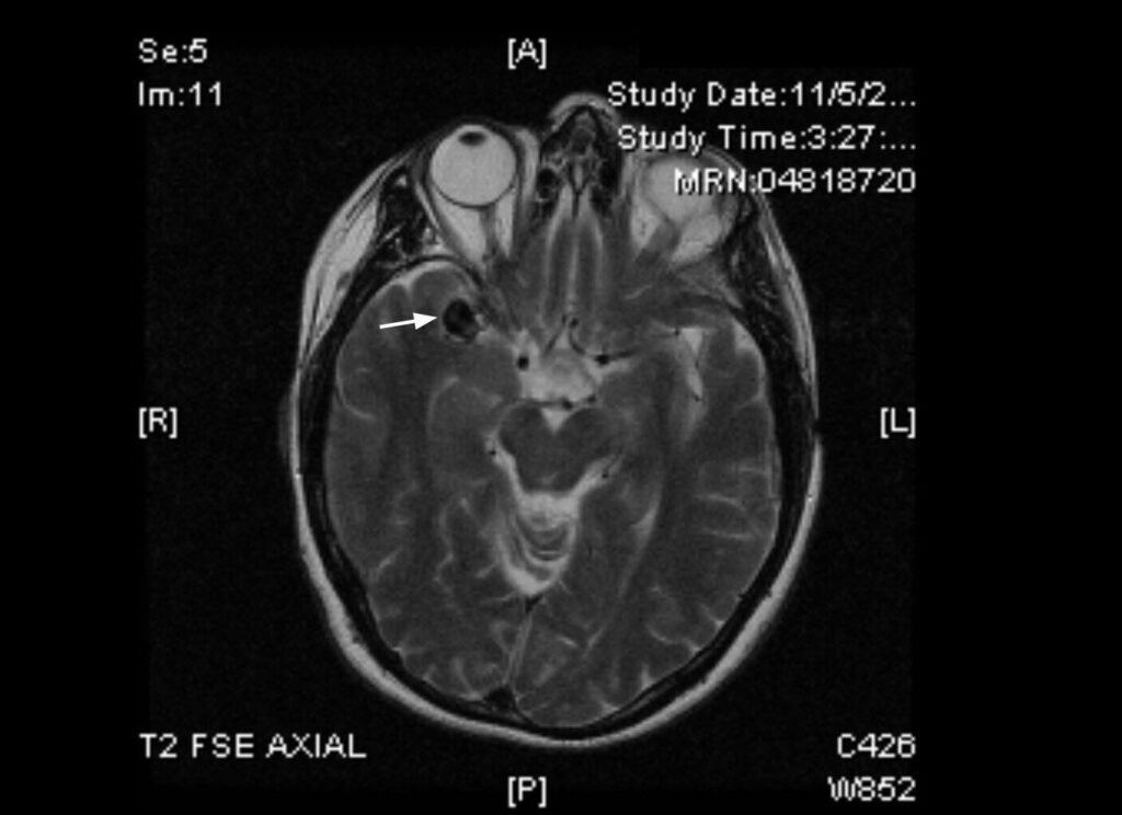
Most unruptured aneurysms cause no symptoms at all. When an unruptured aneurysm does cause symptoms, they may include a dilated pupil, drooping of one eye (ptosis), double vision, or vision changes. These symptoms should be evaluated promptly.
How Is an Aneurysm Diagnosed?
If an aneurysm is suspected — whether due to symptoms, a family history, or an incidental finding on another scan — Dr. Friedlander will use advanced imaging to get a complete picture of its size, shape, and exact location.
Common imaging tests include:
- CT angiography (CTA): A fast, non-invasive scan that uses X-rays and contrast dye to create detailed images of the blood vessels in your brain.
- Cerebral angiogram: Considered the gold standard for evaluating aneurysms, this test involves a thin tube (catheter) inserted into an artery, usually in the wrist or groin, which guides contrast dye to the brain vessels. It provides the clearest, most detailed images of blood flow.
- 3D reconstruction imaging: Using the data from an angiogram, Dr. Friedlander’s team can generate a three-dimensional model of the aneurysm and surrounding vessels.
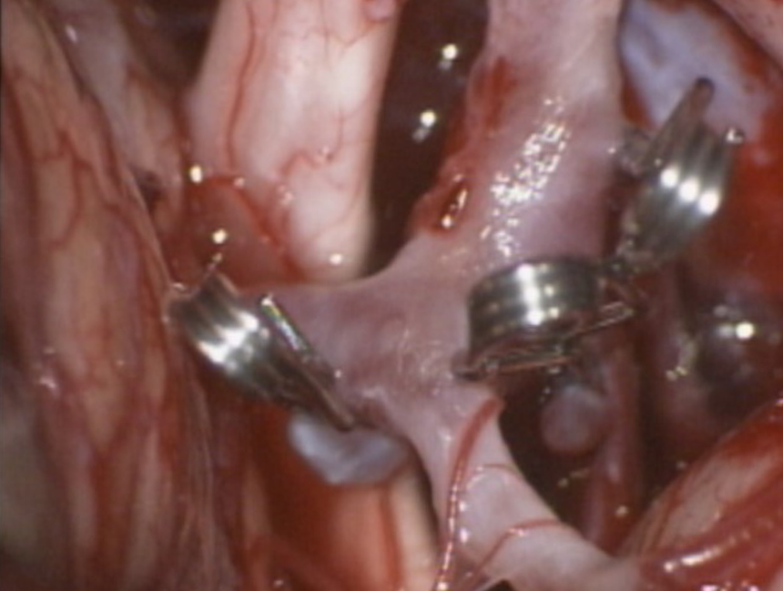
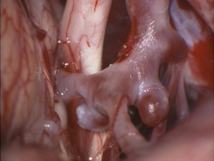

Before and after surgical clipping of three cerebral aneurysms.
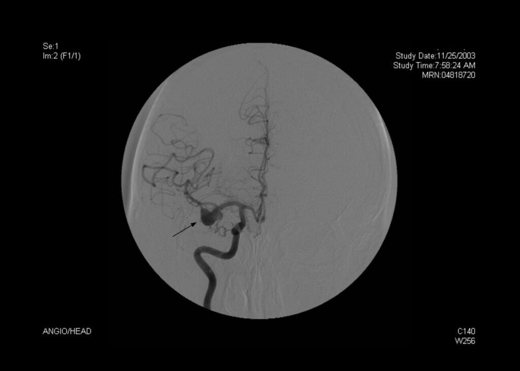
A conventional angiogram showing blood flow through the brain’s vessels and the location of the aneurysm.
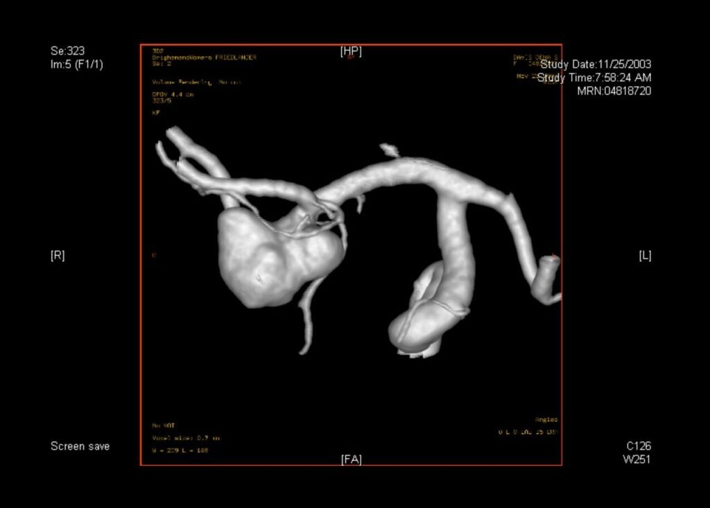
A 3D reconstruction allows Dr. Friedlander to study the aneurysm from every angle before surgery.
Management Decision
After the relevant clinical and imaging information has been obtained, careful analysis is required to determine the most appropriate management strategy. The first and most important question is whether the aneurysm should be treated. This decision is individualized and depends on a thoughtful assessment of both aneurysm-specific and patient-specific factors.
The key consideration is whether the estimated lifetime risk of rupture exceeds the risk of treatment. Rupture risk can be estimated on an annual basis based on aneurysm size, shape, and location, but that estimate must be interpreted in the context of the patient’s age, overall health, and long-term outlook. It must also be weighed against the risks of intervention and the likelihood of achieving a durable result. Thoughtful deliberation about whether to treat is therefore the essential first step.
When treatment is deemed appropriate, the next decision is how to proceed. One option is open microsurgical clipping through a craniotomy, a well-established treatment that Dr. Friedlander performs. Another is endovascular treatment, performed from within the blood vessels, which is less invasive. Each approach has specific advantages and limitations, and the choice must be tailored to the individual patient and aneurysm.
Dr. Friedlander collaborates closely with endovascular specialists to help patients understand their options and make the most informed treatment decision.
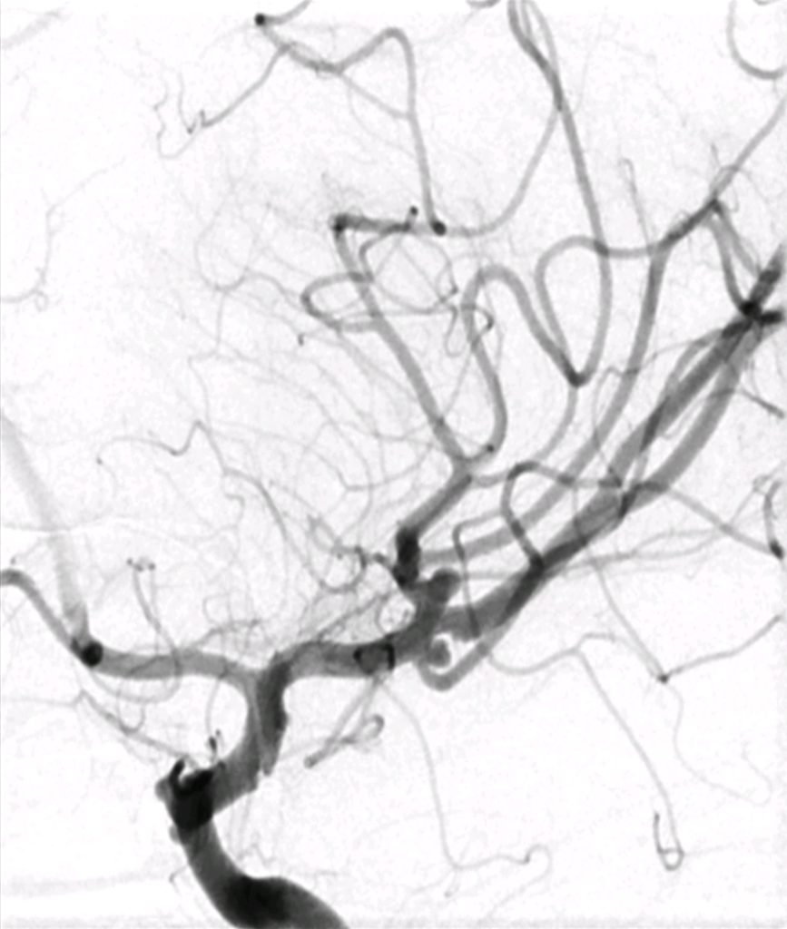
Surgical Clipping: How It Works
Dr. Friedlander has experience surgically treating hundreds of aneurysms.
Before surgery: You will meet with Dr. Friedlander to review your imaging, discuss your treatment options, and if surgery is selected, then develop a plan tailored to your specific anatomy and overall health.
During surgery: Under general anesthesia, Dr. Friedlander makes a small opening in the skull (called a craniotomy) to access the area of the brain where the aneurysm is located. Using a surgical microscope, he carefully navigates to the aneurysm using specialized techniques to minimize brain manipulation. A small titanium clip — roughly the size of a fingernail — is then placed at the base of the aneurysm, sealing it off from the normal blood vessel. The clip remains permanently in place and does not set off metal detectors.
After the clip is placed: Blood no longer enters the aneurysm, providing durable protection against future rupture when complete occlusion is achieved. The surrounding blood vessels continue to function normally.
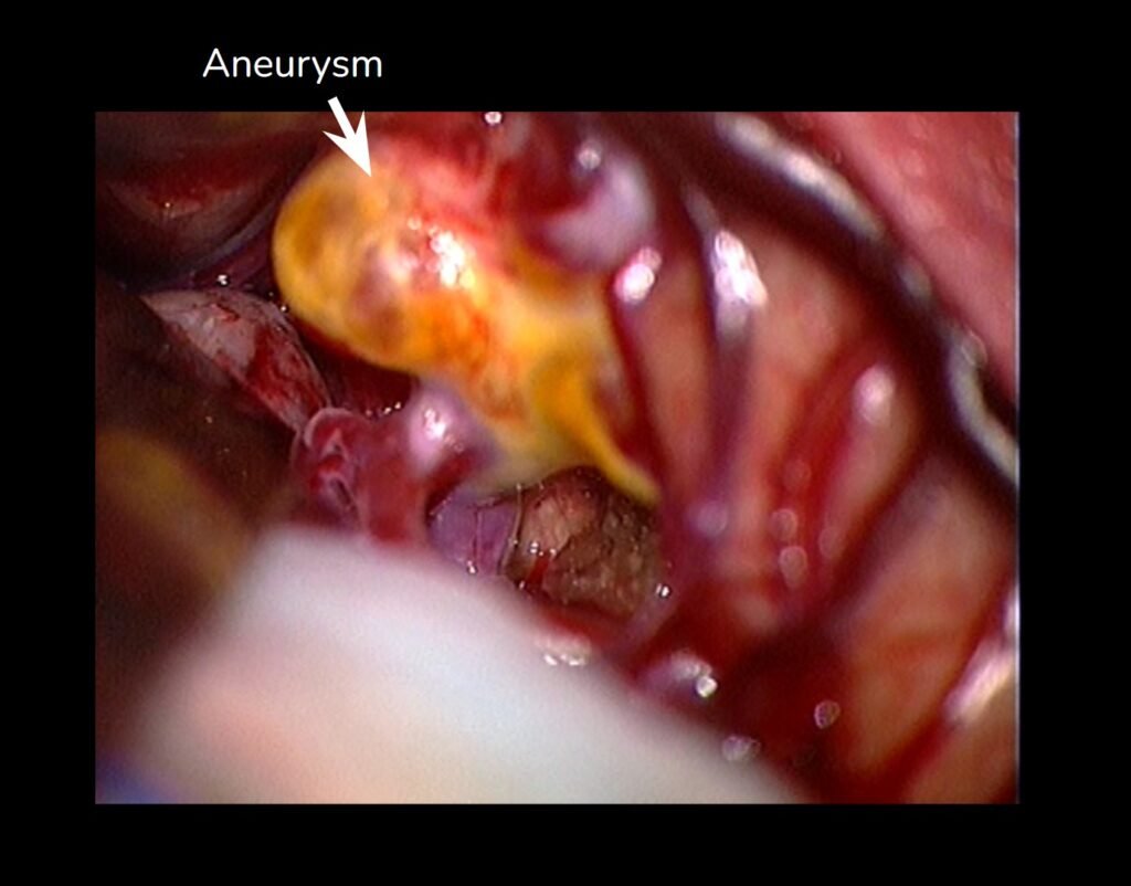
The aneurysm as seen through the surgical microscope during the operation.
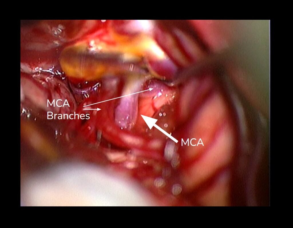
The middle cerebral artery (MCA) and its branches — the blood vessels near the aneurysm — are carefully preserved.
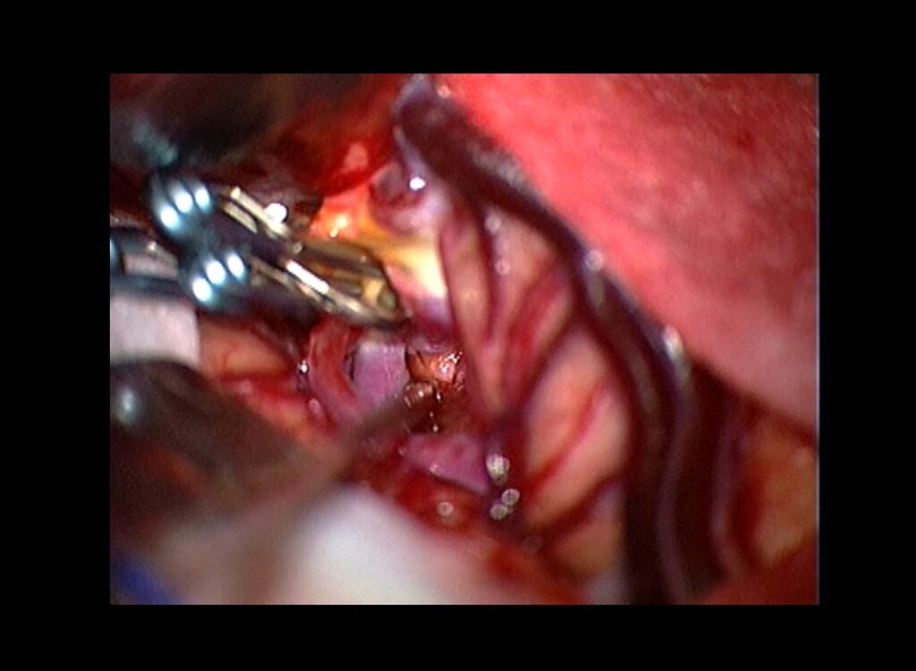
The same area after the titanium clip has been applied, closing off the aneurysm.
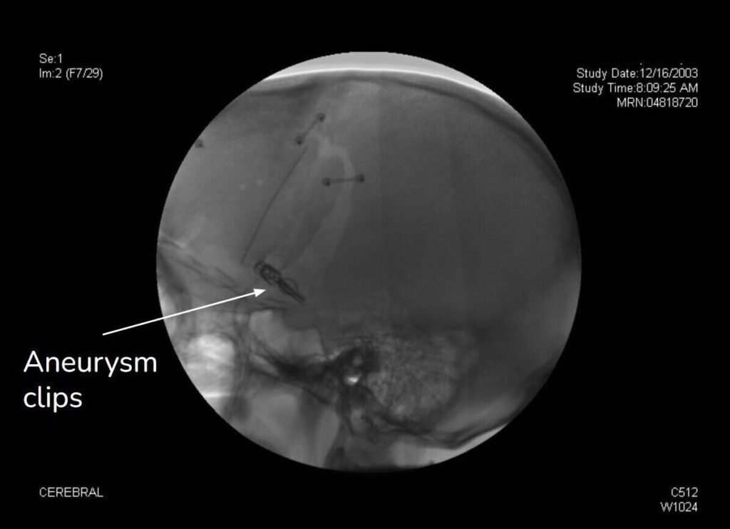
A post-operative X-ray showing the titanium clip in place. The clip is permanent and causes no discomfort.
What to Expect After Surgery
Most patients stay in the hospital for 2 to 3 days following surgical clipping. The recovery experience varies depending on whether the aneurysm had ruptured prior to surgery, but the majority of patients treated for unruptured aneurysms return to normal activities within 2 to 4 weeks.
Confirming success: Before you leave the hospital, imaging is performed to confirm that the aneurysm has been completely and safely clipped, and that blood flow to the surrounding brain is normal.
Long-term outlook: Once clipped, an aneurysm is considered durably treated. Dr. Friedlander’s team will continue to follow your care over time, but the vast majority of patients never experience recurrence.
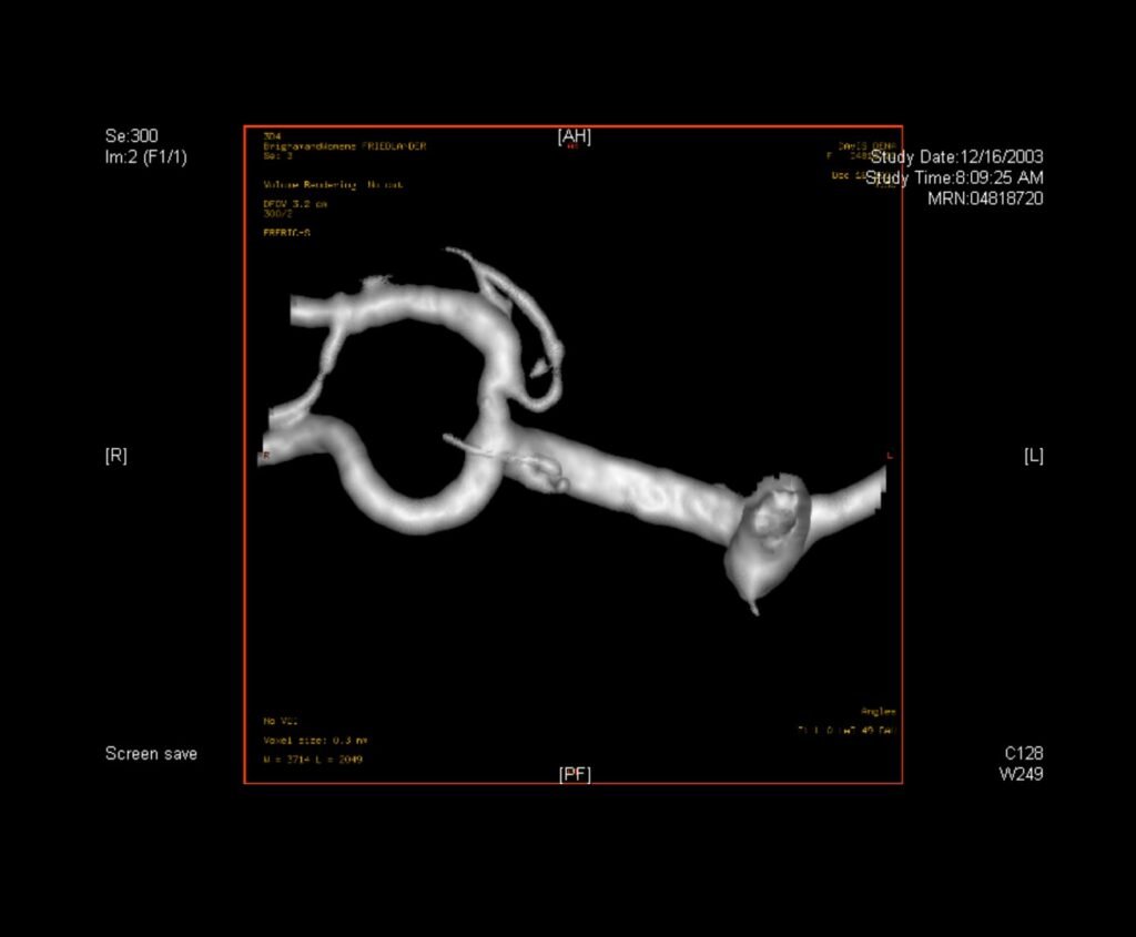
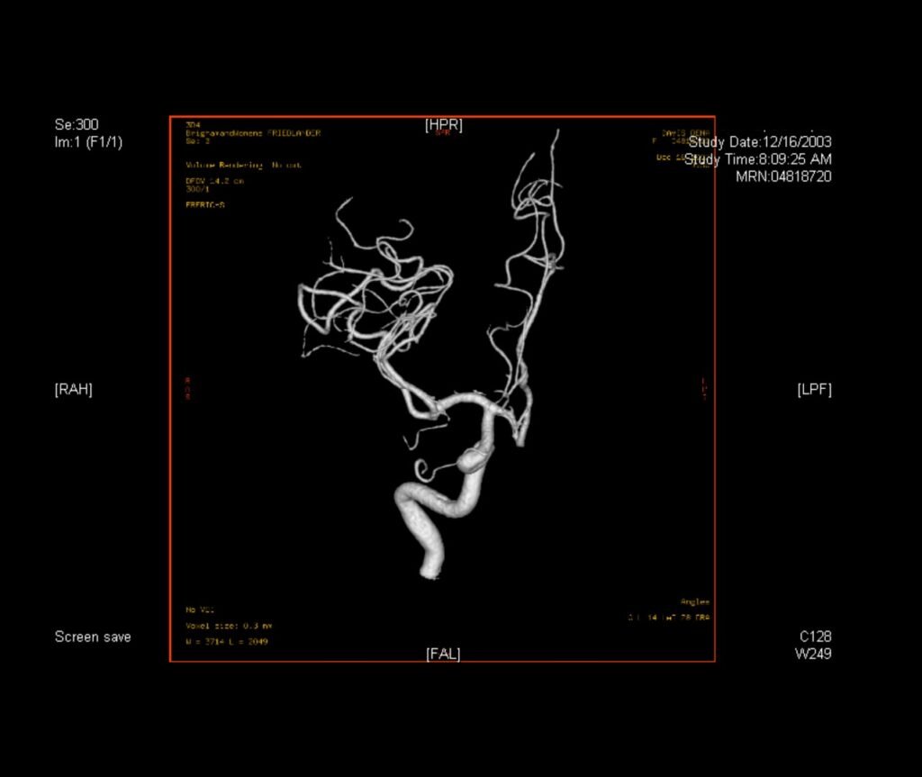
A post-operative 3D angiogram confirming the aneurysm is gone and the blood vessel is fully intact.

Request a Consultation with
Dr. Friedlander
dr.rf@iss.org
Phone
845·764·4353
Address