
Welcome to the
Friedlander Chiari Center
The Chiari Center, directed by Robert M. Friedlander, MD, MA, provides highly specialized evaluation and treatment for adults with Chiari malformation. The Center serves as a national referral destination for patients seeking experienced, evidence-based care for complex, atypical, or previously treated cases.

A National Leader in Adult Chiari Malformation
The Friedlander Chiari Center, directed by Robert M. Friedlander, MD, MA, specializes in the evaluation and surgical treatment of adult Chiari I malformation. With experience evaluating several thousand patients and performing approximately 700 adult Chiari operations, Dr. Friedlander provides individualized Chiari decompression surgery, revision surgery after previous decompression, and specialized second opinions for complex cases.
Located in White Plains, New York, the Center welcomes patients from throughout the United States and abroad. Dr. Friedlander’s surgical philosophy emphasizes restoring normal cerebrospinal fluid flow, relieving neurological compression, and preserving normal anatomy whenever possible.
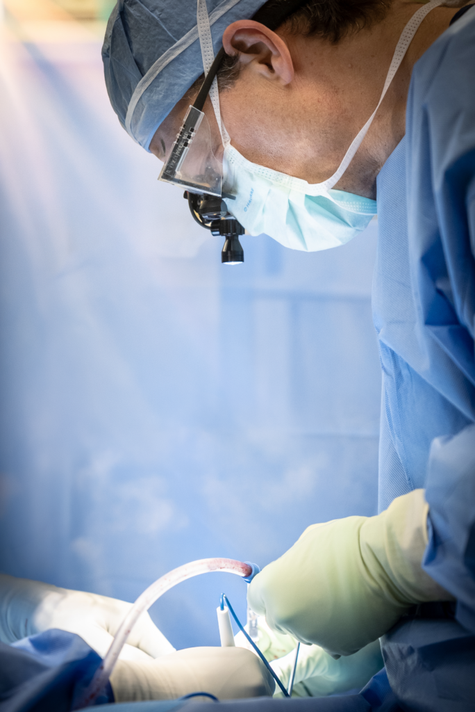
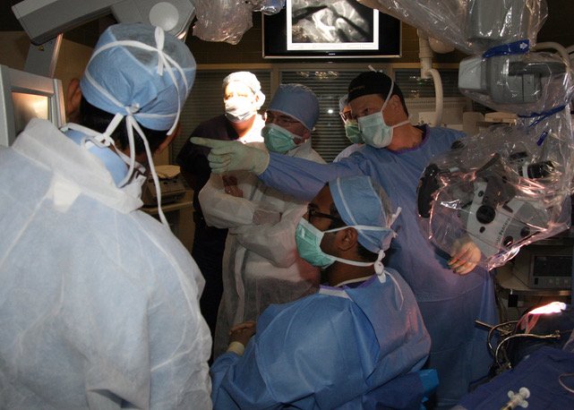
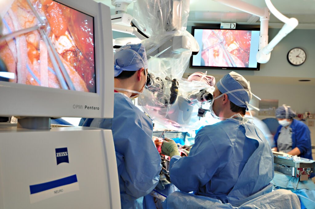
Surgical images are shown below for educational purposes.
A Rigorous Diagnostic Framework
The Center emphasizes precise diagnosis. Not all radiographic tonsillar descent represents symptomatic Chiari malformation, and not all symptoms attributed to Chiari originate from it.
Each evaluation includes:
- Comprehensive neurological assessment
- Detailed imaging review, including CSF flow analysis when indicated
- Assessment for craniocervical instability
- Consideration of alternative or coexisting diagnoses
- Individualized determination of surgical candidacy
Careful differentiation between incidental findings and clinically significant pathology is central to treatment planning, particularly for patients being considered for revision surgery.
Chiari Decompression Surgery: An Individualized Surgical Approach
When surgery is indicated for adult Chiari I malformation, Dr. Robert M. Friedlander’s surgical approach focuses on restoring normal cerebrospinal fluid (CSF) flow, relieving compression of the brainstem and upper spinal cord, and preserving as much normal anatomy as possible.
With experience performing approximately 700 adult Chiari operations, including complex and revision Chiari decompression surgery, Dr. Friedlander tailors each procedure to the patient’s anatomy, symptoms, imaging findings, and CSF flow abnormalities.
His guiding philosophy is to achieve an effective decompression while minimizing unnecessary disruption of the surrounding bone, muscles, and ligaments. The objective is not simply to create more space, but to correct the underlying physiological problem while preserving the structural integrity of the craniocervical junction.
Tailored C1 Laminectomy: Preserving the C1 Arch When Possible
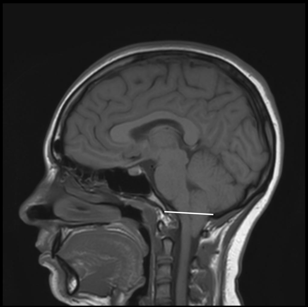
An important consideration in Chiari decompression surgery is whether—and how much—of the posterior arch of C1, the first cervical vertebra, must be removed to relieve crowding at the foramen magnum.
Rather than routinely removing the entire posterior C1 arch, Dr. Friedlander tailors the extent of bone removal to each patient’s anatomy. In selected patients, a limited C1 laminotomy, involving removal of only the superior portion of the posterior arch, can provide adequate decompression while preserving the continuity and structural integrity of the remaining C1 arch.
In other patients, particularly when the cerebellar tonsils extend farther into the upper cervical spinal canal, a more extensive or complete C1 laminectomy may be necessary.
This individualized, anatomy-preserving approach also emphasizes limiting muscular dissection and preserving ligamentous attachments whenever feasible.
The rationale is that minimizing unnecessary disruption of normal supporting structures may help preserve craniocervical stability and reduce postoperative discomfort. Although these potential benefits have not been conclusively demonstrated for this particular C1 technique, preservation of normal anatomy remains an important surgical principle.
The goal is always the same: achieve sufficient decompression without removing more bone or disturbing more supporting tissue than necessary.
Restoring CSF Flow and Relieving Brainstem and Spinal Cord Compression
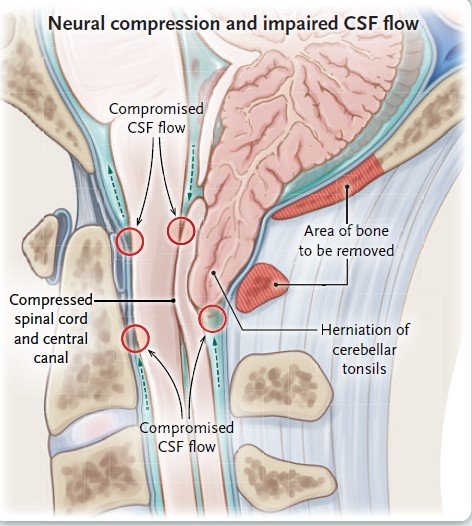
The purpose of Chiari decompression surgery extends beyond enlarging the bony opening at the base of the skull. Cerebellar tonsillar crowding can obstruct cerebrospinal fluid circulation at the foramen magnum and contribute to compression of the brainstem and upper cervical spinal cord.
Dr. Friedlander’s surgical approach emphasizes restoration of unobstructed CSF flow and relief of neurological compression.
Depending on the individual patient’s anatomy and operative findings, techniques may include posterior fossa decompression, dural opening with expansile watertight duraplasty, microsurgical release of arachnoid adhesions, and reduction of crowded cerebellar tonsils.
Microscopic Evaluation of the Foramen of Magendie
A particularly important aspect of Dr. Friedlander’s intradural surgical approach is careful microscopic examination of the fourth ventricle outlet, known as the foramen of Magendie.
In some patients with Chiari malformation, thin arachnoid membranes or adhesions obstruct the outlet of the fourth ventricle, contributing to impaired CSF circulation. These obstructions may not be apparent on MRI and may only become evident once the fourth ventricle is directly evaluated.
Dr. Friedlander considers careful inspection of this region important. When an obstructing membrane or adhesion is identified, it may be released microsurgically when appropriate and safe to do so, helping restore the passage of CSF from the fourth ventricle into the surrounding subarachnoid space.
This attention to the fourth ventricular outlet reflects a central principle of his surgical approach: successful decompression requires addressing the actual sources of CSF flow obstruction, not simply removing bone.
The extent of bone removal, dural reconstruction, and intradural intervention is individualized to achieve adequate decompression while limiting unnecessary surgical manipulation.
Revision Chiari Surgery: Persistent or Recurrent Symptoms After Previous Decompression
Dr. Friedlander has extensive experience evaluating adults who continue to experience symptoms after previous Chiari decompression surgery or whose symptoms return following an initial period of improvement.
Patients seeking revision Chiari surgery or a second opinion after an initial decompression often have persistent headaches, neurological symptoms, impaired CSF flow, or syringomyelia (a fluid-filled cavity within the spinal cord).
However, persistent symptoms do not necessarily mean that the original surgery was inadequate, nor do they automatically indicate that repeat surgery is needed.
A comprehensive evaluation may include review of the original operative report, comparison of preoperative and postoperative MRI studies, assessment of CSF flow when indicated, and consideration of alternative or coexisting causes of symptoms.
When revision Chiari decompression is appropriate, the procedure is directed toward the specific underlying abnormality. Potentially correctable issues include:
- Need for greater decompression, particularly at the level of the foramen magnum
- Persistent obstruction of CSF flow
- Arachnoid scarring or adhesions
- Membranous obstruction of the fourth ventricular outlet (foramen of Magendie)
- Revision of the previous dural reconstruction
- Intradural exploration after prior bone-only decompression
Revision surgery may involve additional bony decompression, microsurgical release of adhesions, restoration of fourth ventricular outflow, or revision of the duraplasty, depending on the operative findings.
The decision to undertake repeat Chiari surgery requires careful judgment, given the likely greater risk of a reoperation. Dr. Friedlander’s goal is to identify a correctable cause of persistent symptoms while avoiding additional procedures that are unlikely to provide meaningful benefit.
Complex Chiari Malformation and Craniocervical Stability
Some adults with Chiari malformation have associated anatomical abnormalities or connective-tissue conditions, including Ehlers-Danlos syndrome, a retroflexed odontoid, or suspected craniocervical instability.
These findings can complicate surgical decision-making and require careful assessment of the relationship between structural abnormalities, neurological symptoms, and CSF flow.
Dr. Friedlander’s approach emphasizes preservation of normal stabilizing structures whenever feasible, with particular attention to the integrity of the C1 arch and the supporting muscles and ligaments of the craniocervical junction.
The presence of an associated anatomical finding does not automatically establish that it is responsible for symptoms or that additional surgery is necessary. Procedures intended to address instability or other structural abnormalities are considered only when supported by the clinical and radiographic findings.
Recovery After Chiari Decompression Surgery
The same philosophy of individualized, anatomically focused surgery extends to postoperative recovery.
By limiting unnecessary bone removal and soft-tissue dissection, Dr. Friedlander seeks to minimize surgical trauma while achieving effective decompression. Careful microsurgical technique and meticulous attention to hemostasis are also important components of his operative approach.
Many patients undergoing uncomplicated primary Chiari decompression are well enough to leave the hospital on the first postoperative day, although hospitalization and recovery vary according to the procedure and individual circumstances.
The ultimate goal is durable clinical improvement, restoration of CSF circulation, and preservation of long-term structural stability, with careful postoperative follow-up to assess symptoms, neurological function, and imaging findings.
How Surgery Addresses the Underlying Cause of Symptoms
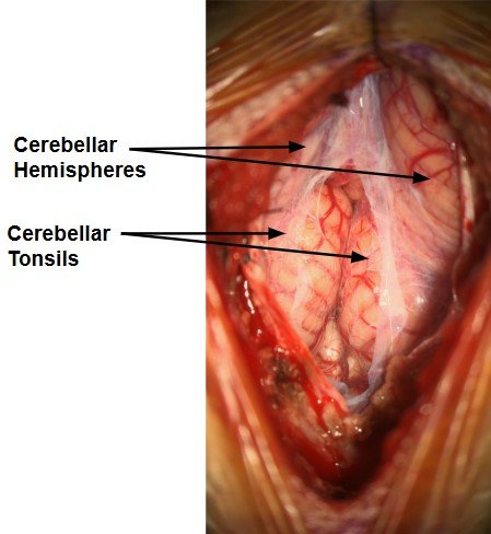
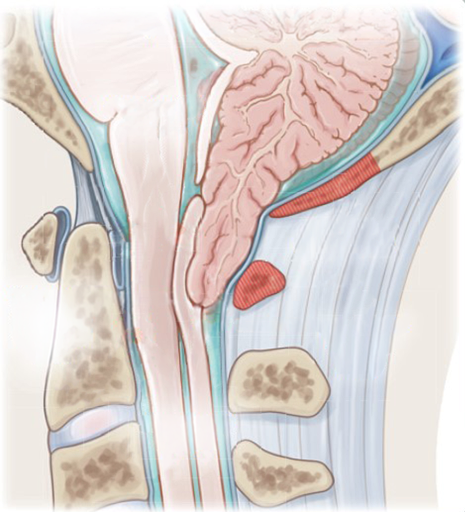
After opening the covering of the brain (the dura), the lower part of the cerebellum (the tonsils) can be seen in a crowded position. This crowding blocks the normal flow of cerebrospinal fluid (CSF), which is a common cause of the characteristic headaches—often worsened by coughing, straining, or other Valsalva maneuvers.
At the same time, the tonsils can place pressure on the upper spinal cord, which may contribute to symptoms such as numbness or tingling in the hands.
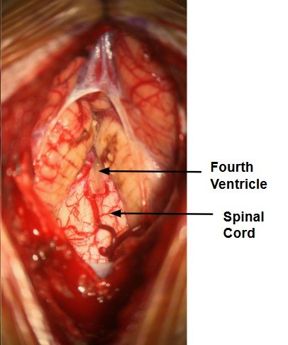
The tonsils are carefully released and reduced in size to relieve both the obstruction to CSF flow and the pressure on the spinal cord. Restoring normal fluid flow from the fourth ventricle and creating space around the spinal cord are key steps aimed at addressing the underlying causes of headaches and hand numbness.
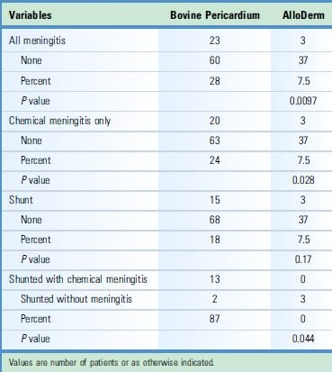
A patch (dural graft) is then placed to create additional space for fluid circulation and to support a watertight closure.
Frequently Asked Questions
Answers to common questions about Chiari malformation evaluation, decompression surgery, and revision surgery. For questions about your specific situation, please contact the office.
No. The extent of C1 removal is determined by the individual’s anatomy, the degree of crowding, and the decompression necessary to restore adequate CSF flow.
Persistent symptoms warrant reassessment of the original diagnosis, previous operation, postoperative imaging, and possible alternative causes. Repeat surgery is not always necessary.
In selected patients, revision decompression may address persistent CSF obstruction, scar tissue, or other correctable structural problems. The decision depends on the findings and anticipated risks and benefits.
These structures support the craniocervical junction. Preserving them when feasible is part of an anatomically focused approach, particularly when structural stability is a concern.
A second opinion can help clarify whether symptoms are attributable to Chiari, whether surgery is appropriate, and which surgical approach best matches the findings.
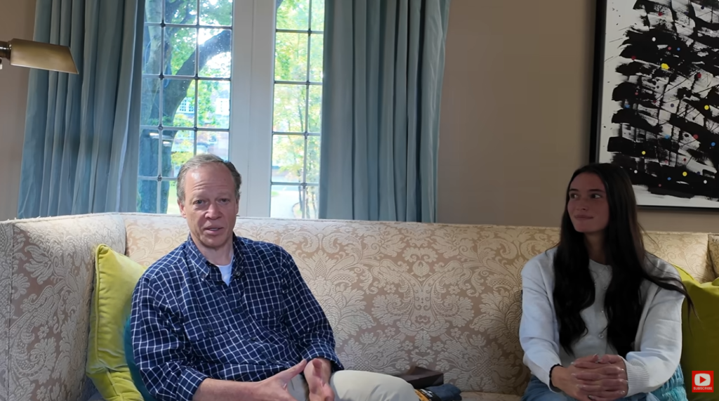
Eva and her family sat down once more to talk with Dr. Robert Friedlander to discuss Eva’s complete recovery. Together, they discuss the realities of neurosurgery, patient care, brain health, and the recovery process for Chiari malformation, drawing on personal experiences to provide insight into this medical field.
Watch now to hear Eva’s story firsthand and gain a deeper understanding of the patient experience, the realities of neurosurgery, and the impact of compassionate, personalized care.
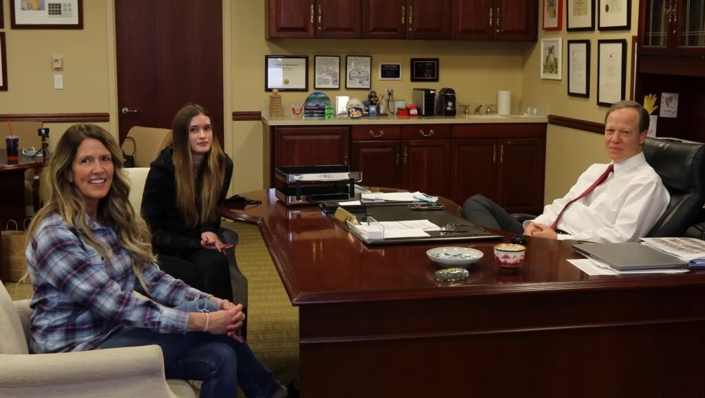
For years, Eva lived with daily headaches, constant pressure in the back of her head, and numbness in her hands and feet—symptoms that began to affect every part of her life. After being diagnosed with Chiari malformation, she came to Dr. Robert Friedlander for specialized care.
Following surgery, Eva experienced immediate relief. Her headaches were gone, the pressure had lifted, and she finally felt like herself again. Watch her story to hear about her journey and recovery firsthand.
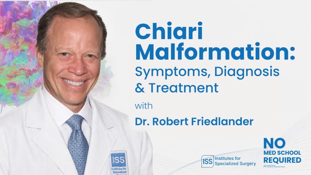
In this in-depth educational interview, Dr. Robert Friedlander, an internationally recognized neurosurgeon who trained at Harvard Medical School and Massachusetts General Hospital, discusses Chiari malformation, its symptoms, diagnosis, treatment options, and surgical considerations.
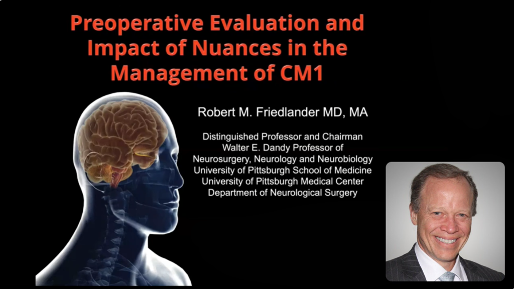
In this presentation for Conquer Chiari’s 2022 A Day of Hope conference, Dr. Robert Friedlander discusses preoperative evaluation and the clinical nuances involved in managing Chiari I malformation.
National Referral Practice
Patients travel from around the nation for evaluation by Dr. Friedlander. Referrals are accepted from neurologists, neurosurgeons, primary care physicians, and directly from patients.
Each case is reviewed with intellectual rigor, surgical experience, and a commitment to clarity. The goal is straightforward: accurate diagnosis, thoughtful surgical judgment, and outcomes grounded in experience and evidence. In evaluating often complex presentations in Chiari patients, there is no substitute for broad experience.
Recent Work
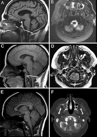
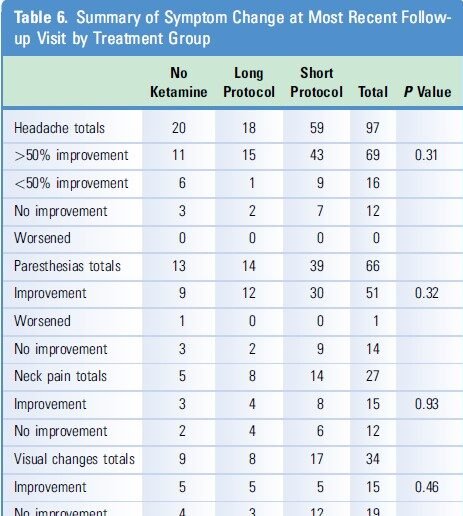
Recent work using formal neuropsychological evaluation has demonstrated that cognitive and affective symptoms—often reported by Chiari patients—can be reversible following expert surgical intervention in appropriately selected individuals. This work has helped reframe the broader neurological impact of Chiari malformation beyond headache and other typical symptoms.

Advancing the Field
Dr. Friedlander authored a single-author review in The New England Journal of Medicine on Chiari malformation and has published research advancing the understanding of both the structural and functional aspects of the disorder.


Request a Consultation with
Dr. Friedlander
dr.rf@iss.org
Phone
212·404·7606
Address